Description
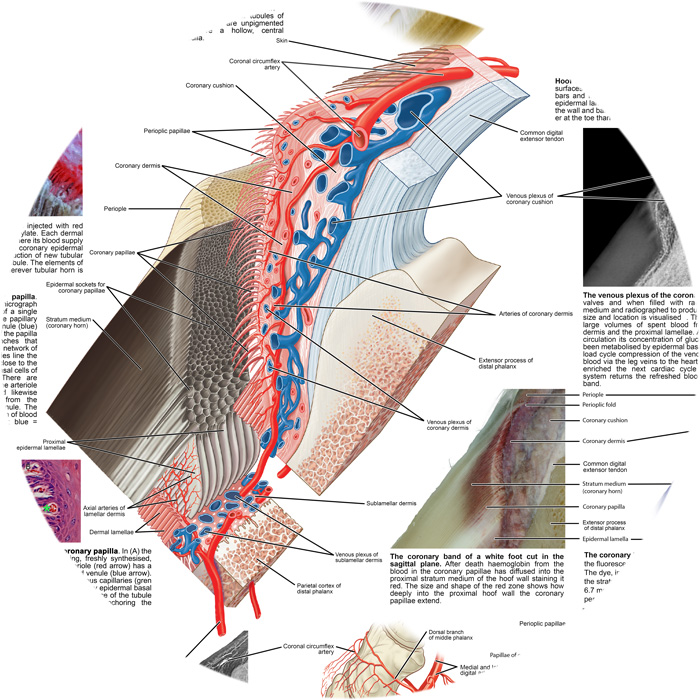
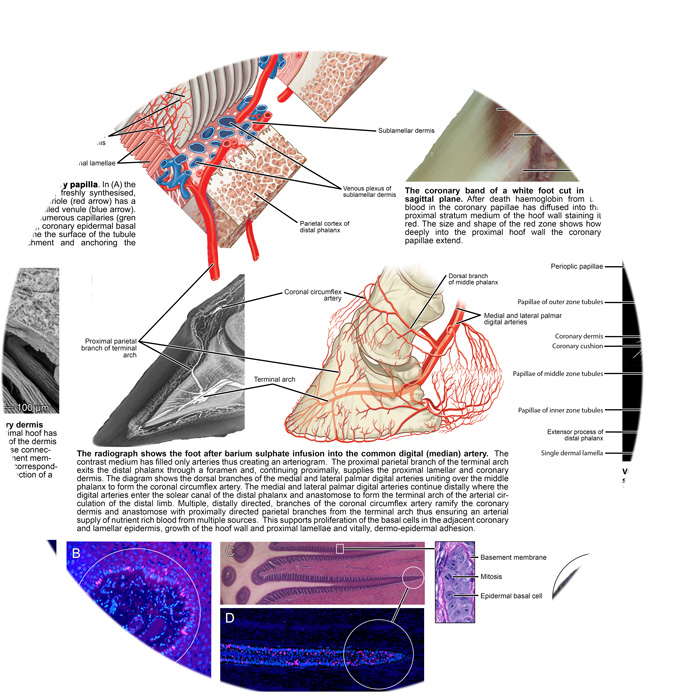
The centre point of each poster is artwork by Richard Tibbitts of www.antbits.com The 3 dimensional artwork was designed by Chris Pollitt to show the anatomy of the proximal hoof wall, the coronary papillae, the proximal dermal and epidermal lamellae and major blood vessels and bones. The dermis and epidermis are separated to illustrate the relationship between these 2 important anatomical compartments. Adjoining the main figure are 24 additional illustrations of proximal hoof wall anatomy, histology, radiology and electron micrography each with its own explanatory caption.
The richly illustrated scientific posters are in-depth study sheets designed to improve knowledge and learning of the equine foot. Ideal for students of equine foot science. Others in the series are; The lamellae and the suspensory apparatus of the distal phalanx; The white line and the terminal papillae; The blood circulation of the equine foot; Laminitis and failure of the suspensory apparatus.
Posters are A1 size only, printed and laminated by The Poster Factory and shipped in tubes.
Reference
The Illustrated Horse’s Foot: A Comprehensive Guide by Christopher C. Pollitt. Elsevier. ISBN: 978-0-
702-04655-1


