Description
from https://3dvetanatomize.com/
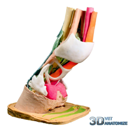
An educational/demonstration tool for students, farriers, trimmers and veterinarians to show what is meant by laminitis, normal range of motion, club foot and broken hoof/pastern axis.
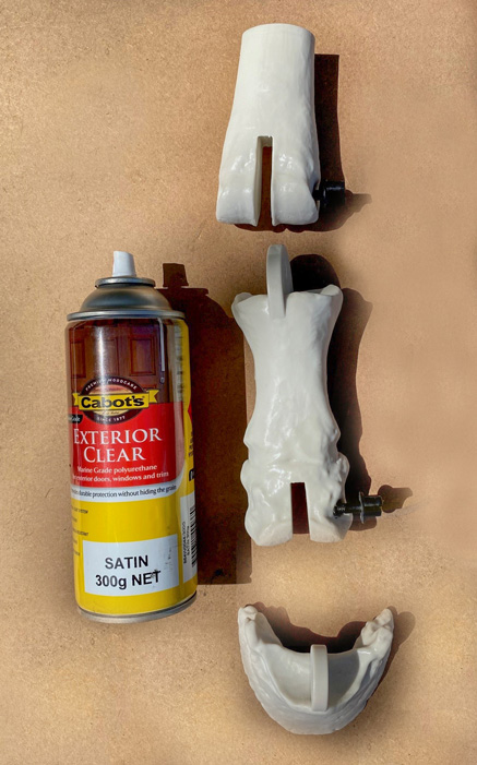
The model consists of 4 bones, 2 hoof capsules (3D printed in durable resin) and 2 flexible dermal inserts (3D printed in flexible polyurethane). The middle and proximal phalanges are fused and treated as a single bone. One side of the hoof capsule is cut away so that contents can be viewed. Two black, nylon thumbscrews are used to lock the articulations in place.
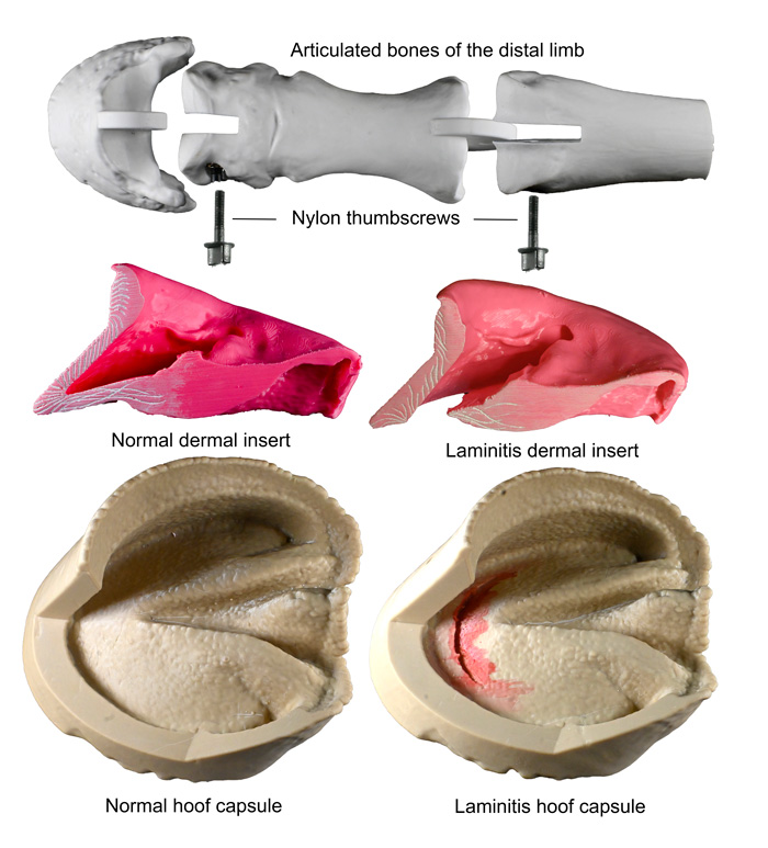
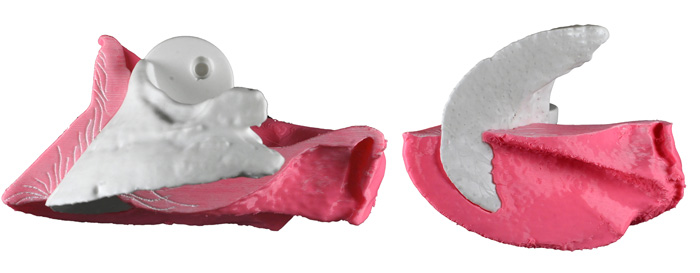
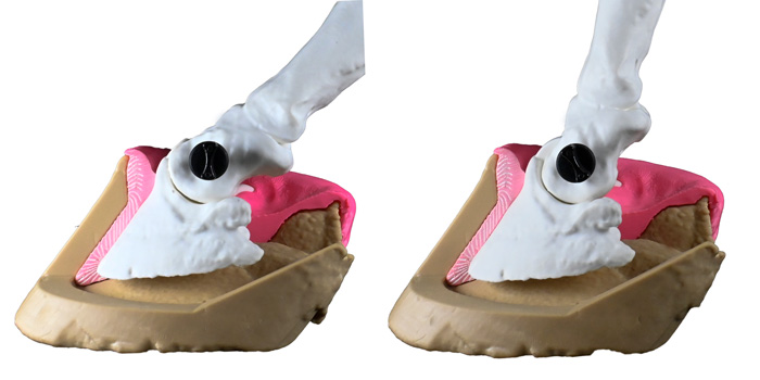
A pink, flexible, polyurethane insert replicates the dermis (corium). Fitting snug around the distal phalanx, it fixes the bone in its correct anatomical position within the hoof capsule. The embossed white markings represent the collagen bundles of the suspensory apparatus of the distal phalanx that suspend the distal phalanx from the lamellae of the inner hoof wall.
Likewise, a similar flexible insert replicates a dermis affected by laminitis. Now the collagen bundles of the suspensory apparatus are no longer connected to the hoof wall lamellae and the distal phalanx has been forced down (foundered) into the hoof capsule, penetrating and crushing the dermis of the dorsal sole.
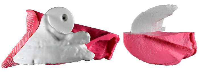
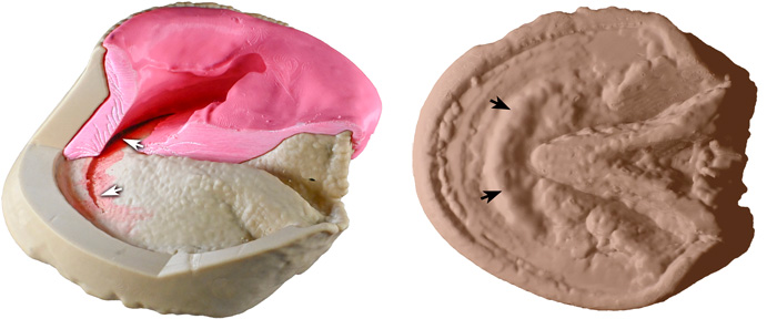
The crushed vascular dermis that is trapped between the semi-circular margin of the distal phalanx and the rigid horn of the sole includes the arterial blood supply of the sole. Thus, sole in the vicinity of the dislocated bone, without adequate arterial blood, undergoes necrosis. This leaves a semi-circular imprint (white arrows) on the inside of the sole and a corresponding bulge (black arrows) on the outside.
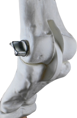
A circular bridle joint (a type of mortise and tenon joint) enables articulation around the centre of rotation of the coffin joint. The mortise (slot) is in the distal end of the middle phalanx and the tenon bisects the proximal distal phalanx thus replicating the ginglymus (hinge) joint of the coffin joint. A nylon thumbscrew screwed tight into a threaded insert in the middle phalanx locks the articulation in the desired position.
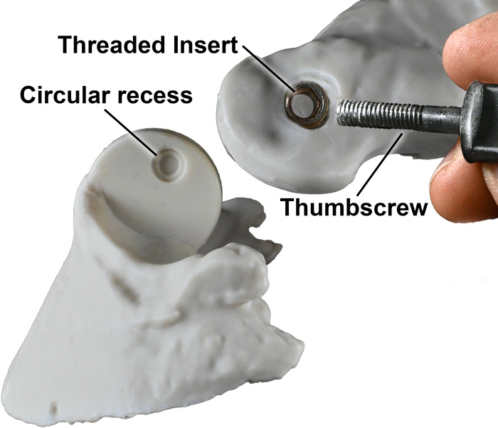
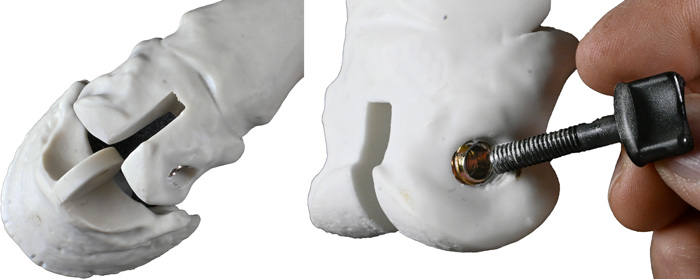
Screwing the thumbscrew into the circular recess in the tenon of the distal phalanx locates the tip of the thumbscrew at the centre of rotation of the joint. Likewise, a tenon embedded in the centre of the proximal phalanx fits into the mortise in the distal metacarpus. Insertion of the thumbscrew locates the circular recess at the centre of rotation and replicates the articulation of the fetlock joint.
When assembled the model can be used to demonstrate the normal range of motion that the joints of the distal limb undergo during the limb cycle – fetlock extension (dorsi-flexion) is shown on the left and fetlock flexion on the right. The coffin joint is flexed in both images.
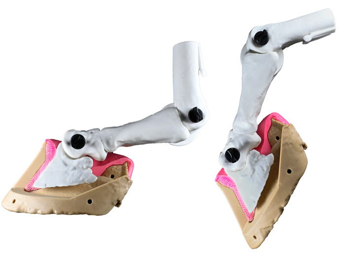
In addition, the model can illustrate the hoof/pastern axis. A broken forward hoof/pastern axis, or club-foot, is shown on the left. A pathological broken back hoof/pastern axis is shown on the right.
The surface of the model is coated with a clear polyurethane finish to seal in resin dust and ensure a long working life.