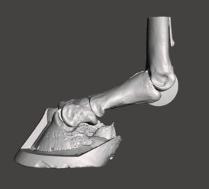
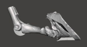
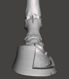
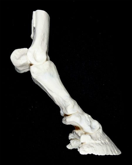
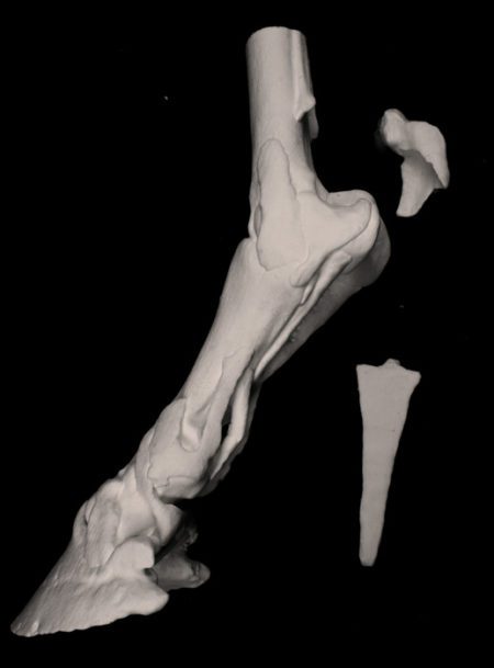
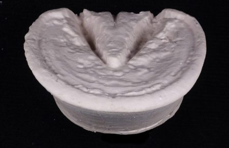
The articulated model of the horse distal limb

$450.00 USD

3DVetAnatomize bring together an international team of veterinary imaging and computer modelling specialists to create unique anatomical replicas of the horse’s distal limb to meet your specific learning, teaching and demonstration needs.

Category:

Description

The articulated model of the horse distal limb

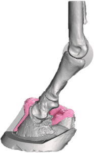
An educational/demonstration tool for students, farriers, trimmers and veterinarians. Show what you mean by club foot, broken back hoof/pastern axis and medial lateral balance.

The model consists of 4 bones and a hoof capsule, 3D printed in white plastic. One side of the hoof capsule is cut away so that contents can be viewed.
A silicone insert replicates the dermis (corium) and fits snugly around the distal phalanx and fixes it in its correct anatomical place.
Articulation around the coffin and fetlock joints is enabled using a system of discs and slots. A disc embedded in the midline of the distal phalanx fits into a reciprocal slot in the middle phalanx thus and replicating the, ginglymus (hinge) joint of the coffin joint.


Likewise, a disc inserted in the third metacarpal (cannon) bone fits into a slot in the proximal phalanx thus replicating the articulation of the fetlock joint.

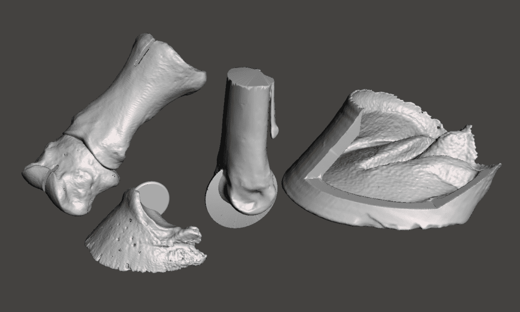
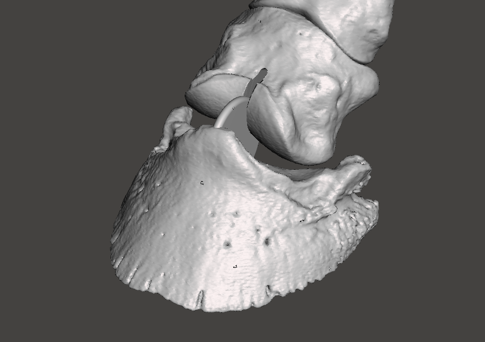
When assembled the model can be used to demonstrate the normal range of motion that the joints of the distal limb undergo during the limb cycle – extension (dorsi-flexion) and flexion.

extension (dorsi-flexion)
flexion
IN ADDITION, THE MODEL IS IDEAL FOR CONVEYING THE CONCEPT OF THE CORRECT HOOF/PASTERN AXIS. A BROKEN FORWARD HOOF PASTERN AXIS, OR CLUB-FOOT IS SHOWN ON THE LEFT. A PATHOLOGICAL BROKEN BACK HOOF PASTERN AXIS IS SHWN ON THE RIGHT.
IN ADDITION AN INCORRECT MEDIAL/LATERAL BALANCE CAN BE SIMULATED TO DEMONSTRATE ITS LIKELY UNDESIRABLE CONSEQUENCES.


Additional information

Weight 0.75 kg
Dimensions 21 × 29 × 12 cm
Format

The basic ‘RE-BUILD’ model, The basic ‘PLUGGED-IN’ model, The ‘ADVANCED’ model